Popular Science Lecture Series - 16
Nuroimaging using fMRI


Venue: Anna Centenary Library,
Kotturpuram
Chennai


January 25, 2020
3:00 pm - 5:30 pm

Co-organised by
Anna Centenary Library
& Tamilnadu Science Forum

About Program


This is part of its efforts to popularize science to the General Public and Students who are pursuing science as their career. TNSF attempt to focus on students on higher science as everyone knows that learning of science at college within the curriculum is not enough to acquire holistic knowledge of science at the appropriate time. Hence, to fill the gap between what students are acquiring through the curriculum and what it is required, TNSF is planning its activities on higher science to students who are pursuing higher education


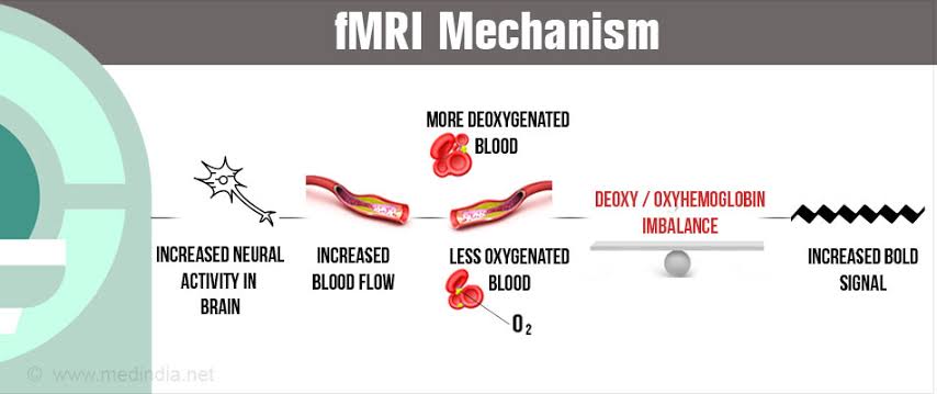
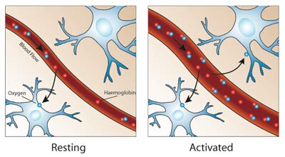
Functional magnetic resonance imaging, or FMRI, works by detecting the changes in blood oxygenation and flow that occur in response to neural activity – when a brain area is more active it consumes more oxygen and to meet this increased demand blood flow increases to the active area. FMRI can be used to produce activation maps showing which parts of the brain are involved in a particular mental process.


How does MRI work?
FMRI is a special type of magnetic scan. The cylindrical tube of an MRI scanner houses a very powerful electro-magnet. A typical research scanner (such as the FMRIB Centre scanner) has a field strength of 3 teslas (T), about 50,000 times greater than the Earth’s field. The magnetic field inside the scanner affects the magnetic nuclei of atoms. Normally atomic nuclei are randomly oriented but under the influence of a magnetic field the nuclei become aligned with the direction of the field. The stronger the field the greater the degree of alignment. When pointing in the same direction, the tiny magnetic signals from individual nuclei add up coherently resulting in a signal that is large enough to measure. In FMRI it is the magnetic signal from hydrogen nuclei in water (H2O) that is detected. The key to MRI is that the signal from hydrogen nuclei varies in strength depending on the surroundings. This provides a means of discriminating between grey matter, white matter and cerebral spinal fluid in structural images of the brain.

காந்த அதிர்வு அலை வரைவு அல்லது M.R.I. ஸ்கேன் (Magnetic Resonance Imaging) என்பது சக்தி வாய்ந்த காந்தத்தின் உதவியுடன் கம்ப்யூட்டர் மூலம் திரையில் காண்பது. காந்த அதிர்வு அலை வரைவு (M.R.I. ஸ்கேன்) உதவியுடன் மூளை, தண்டுவடம், நரம்பு, எலும்பு சம்பந்தமான நோய்களின் தன்மைகளை கண்டறியமுடியும். ஆனால் அடிக்கடி இந்தச் சோதனைகள் செய்தால் பாதிப்பு ஏற்படும். அல்ட்ரா சவுண்ட் ஸ்கேனில் பக்க விளைவுகள் இல்லை. இம்முறை புரோட்டான் படமாக்க முறை (proton imaging ) என்றும் அறியப்படுகிறது.காரணம் புரோட்டான்களே படிமங்களைப் பெற முக்கிய பங்களிக்கிறது. எம்.ஆர்.ஐ எந்திரத்தின் மூலம் உடற்பிரச்சனைகளை நாம் மிகத் துல்லியமாக கண்டுபிடிக்கலாம். காந்த ஒத்திசைவு படமெடுத்தல் என்பதை ஆங்கிலத்தில் மேக்னடிக் ரெசொனென்ஷ் இமேஜிங்க் என்று கூறுவதால் இந்த இயந்திரத்தை எம்.ஆர்.ஐ என்று அழைக்கிறோம். இதை மருத்துவமனையில் நோயாளிகளை ஸ்கேன் செய்ய பயன்படுத்துகிறார்கள். உடலிலுள்ள சில காயங்களை வெறுங்கண்ணால் காண முடியாது. அவ்வாறு கண்டுபிடிக்க முடியாத காயங்களை மிகைப்படுத்தி பார்க்க எம்.ஆர்.ஐ ஸ்கேன் உதவுகிறது. எம்.ஆர்.ஐ மூலம் மூளை கட்டி, மூட்டு வலிகள் மற்றும் மாரடைப்பு போன்ற பெரும் வியாதிகளையும் கூட நம்மால் கண்டுபிடிக்க முடியும். எனவே சில கண்டுபிடிக்க முடியாத தீவிர நோய்களைக் கண்டுபிடிக்க எம்.ஆர்.ஐ பேருதவி புரிகிறது. காந்த அதிர்வு அலை வரைவு அணுக்கரு காந்த ஒத்திசைவைப் பயன்படுத்தியே கணனியில் படங்களைத் தோற்றுவிக்கின்றன.

Program Schedule

Time: 3:00 pm to 5:30 pm - January 25th, 2020

Registration: 2:30 pm onwards

3:00 pm - 4:00 pm

Dr. Raghuram Chetty
Professor,
Department of Chemical Engineering, Indian Institute of Technology - Madras

Fundamentals and applications of lithium-ion batteries

4:00 pm -5:00 pm

Dr. Muali Murugavel
Assistant Professor,
Department of Chemical Engineering, Indian Institute of Technology - Madras

What next in batteries for electric vehicles - A look into the Indian context!

5:00 pm - 5:30 pm

Question & Answer

Speakers

Dr. Raghuram Chetty

Dr. Raghuram Chetty

Dr. Raghuram Chetty is a Professor at the Department of Chemical Engineering, Indian Institute of Technology Madras, Chennai. His area of interest are Electrocatalysis, Fuel cells, Carbon nanotubes. He has his post doctoral assignments with Laboratory of Industrial Chemistry, Ruhr University Bochum, Germany[2007-08] and Chemical Engineering and Advanced Materials, Newcastle University, United Kingdom[2005-07]. He is part of IIT-M since 2009.

Aravind Kumar Chandiran Ph.D

Aravind Kumar Chandiran Ph.D

Aravind Kumar Chandiran Ph.D, is Assistant Professor at Department of Chemical Engineering, Indian Institute of Technology Madras, Chennai. His are of interest are Solar cells, Solar water splitting, Carbon dioxide reduction, Photoconductivity and Oxide semiconductors. His doctoral degree is from Swiss Federal Institute of Technology (EPFL), Lausanne, Switzerland[2010-2013]. He was affliate postdoc in Molecular Foundry, Lawrence Berkeley National Laboratory, USA. His Postdoc was with Long Group, Department of Chemistry University of California, Berkeley, USA.

Venue

Anna Centenary Library,
Kotturpuram
Chennai

Call

+91 9176512565

Email us

tamilnadutnsfchennai@gmail.com